Medical school is an intellectual crucible, demanding not just the rote memorization of countless facts, but the deep, intuitive understanding of complex biological systems. Among the foundational pillars of this grueling education is human gross anatomy. For centuries, the study of the human body was a painstaking process of poring over heavy, two-dimensional textbooks and participating in strictly timed, highly stressful cadaver dissections. While these traditional methods remain respected cornerstones of medical pedagogy, they possess inherent limitations that can hinder a student’s spatial comprehension. Textbooks force the brain to perform a complex cognitive leap, translating flat, stylized images into intricate, three-dimensional living structures—a leap that can easily lead to misinterpretations of spatial relationships.
Cadaver laboratories, while undeniably invaluable for understanding actual tissue texture and biological variation, are restricted by limited access hours, ethical constraints, and the irreversible nature of physical dissection. Once a nerve is severed or a vessel is retracted, the specific anatomical relationship is destroyed, and it cannot be reconstructed for a peer to study. Consequently, there has been a profound pedagogical shift in how future physicians master the intricacies of the human form. Today, high-fidelity replicas serve as the crucial bridge between abstract theory and clinical reality. This comprehensive guide explores the evolution, pedagogical benefits, system-specific applications, and purchasing strategies surrounding these indispensable educational tools.
The Historical Context of Anatomical Education
To truly appreciate the immense value of modern educational tools, one must understand the fraught and fascinating history of anatomical study. In the ancient world, scholars like the Greek physician Galen were legally and religiously barred from dissecting human remains. They were forced to extrapolate human anatomy from the dissection of Barbary macaques and pigs, leading to centuries of profound anatomical misconceptions that were treated as gospel. It was not until the Renaissance, fueled by the relentless curiosity of pioneers like Andreas Vesalius and artists like Leonardo da Vinci, that direct human dissection became the undisputed gold standard for understanding human physiology.
However, preserving human remains was a near-impossible feat before the advent of modern chemical embalming techniques in the late 19th century. Bodies decomposed rapidly, making prolonged study impossible. Furthermore, the demand for bodies by medical schools in the 18th and 19th centuries vastly outstripped the legal supply, leading to the grim era of “body snatchers” and grave robbing. To combat these logistical and ethical nightmares, the first true anatomical teaching aids were created. Master artisans in Italy collaborated with leading physicians to sculpt breathtakingly lifeli ke, life-sized models out of colored beeswax. These wax figures allowed students to study the superficial veins and deep musculature without the immediate need for a decomposing cadaver.
As medical education became highly formalized in the 20th century, the demand for durable, mass-produced study materials surged. Wax was notoriously fragile and highly sensitive to temperature changes, prompting a shift toward papier-mâché and, eventually, durable plastics and resins. Today, the landscape of medical education is facing new, modern challenges. The financial cost of maintaining modern cadaver labs with specialized ventilation is astronomical, and institutions are seeking ways to minimize student exposure to toxic embalming chemicals like formaldehyde and phenol. The evolution of anatomical replicas is not merely a story of technological advancement; it is a necessary, practical response to the logistical and health demands of modern medical training.
The Pedagogical Advantages of Tactile Learning
The leap from understanding an anatomical concept on a textbook page to recognizing that same structure in a living, breathing patient in the surgical theater is perhaps the most difficult transition in a young doctor’s career. This is precisely where the physical interaction with highly detailed anatomy models for medical students becomes a game-changer. These physical tools offer several profound pedagogical advantages that traditional media simply cannot replicate.
First and foremost is the undeniable benefit of haptic feedback and kinesthetic learning. The human brain is evolutionarily hardwired to learn through touch and physical manipulation. When a student physically holds a replica of a sphenoid bone, their brain actively processes the weight, the texture, and the complex, bat-like shape in a way that passive visual observation cannot achieve. They can trace the path of the optic nerve through the optic canal with their fingers, or physically feel the depth of the sella turcica, creating a strong kinesthetic memory. This “muscle memory” is far easier to recall under the intense psychological pressure of a practical board exam or during a high-stakes surgical rotation.
Secondly, these tools foster true three-dimensional spatial awareness and a mastery of directional terminology. The human body is a marvel of compact engineering; structures do not exist in isolation but are intricately layered, interwoven, and packed tightly together. A classic two-dimensional atlas, such as Netter’s Atlas of Human Anatomy, is beautiful and highly detailed, but it fundamentally struggles to convey depth. Students often struggle to understand the relationship between deep and superficial fascia, or how the recurrent laryngeal nerve loops under the aortic arch. By physically taking a model apart and putting it back together, students develop an intuitive, three-dimensional mental map of the body. They can clearly see what is anterior versus posterior, medial versus lateral, and proximal versus distal.
Furthermore, the reusability of these replicas provides an environment designed for infinite repetition and mistake-driven learning without consequence. In a gross anatomy lab, if a student accidentally cuts through the phrenic nerve while attempting to isolate the heart, the specimen is permanently altered. Physical models allow students to dissect and reconstruct complex regions, such as the intricate flexor muscles of the forearm or the delicate bony structures of the inner ear, dozens of times until the spatial relationships become second nature. Finally, the sheer convenience and accessibility of having a desktop model available for a 2:00 AM study session, right before a major midterm, cannot be overstated.
See also: Andrew Pudewa Net Worth: How Much Is the Educator Worth?
A Deep Dive into System-Specific Replicas
The structural diversity of the human body requires a diverse array of teaching tools. Depending on the specific block or bodily system a medical cohort is currently studying, distinctly different types of replicas are required to maximize learning efficiency. Let us explore the most critical categories of models.
Osteology: The Skeletal Foundation
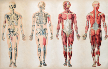
The skeletal system provides the literal and figurative framework for all other anatomical studies. Without a rock-solid understanding of osteology, it is entirely impossible to master muscle origins and insertions, or the complex anatomical pathways of major nerves and blood vessels. Articulated skeletons—fully assembled, life-size replicas hanging from a stand—are vital for understanding joint mechanics, human posture, and the structural relationship between the axial skeleton (skull, spine, ribcage) and the appendicular skeleton (limbs). However, for a first-year student, a disarticulated skeleton is often considered the most valuable tool. By handling individual, loose bones, students can easily identify specific landmarks: the superior and inferior articular processes, the transverse foramina of cervical vertebrae, and the subtle morphological differences between the tibia and the fibula.
Myology and the Muscular System
Understanding the muscular system requires models that explicitly demonstrate the concept of layering. High-quality myology replicas are heavily modular. They allow the student to physically peel away the superficial muscles, such as the massive trapezius and latissimus dorsi of the back, to reveal the deeper musculature like the rhomboids, levator scapulae, and erector spinae group. The most advanced, premium models also include meticulously hand-painted markings detailing the exact origin (typically painted in red) and insertion (painted in blue) points of every single muscle directly on the underlying bone. This visual coding forces the student to understand the biomechanical leverage each muscle exerts on the skeleton when it contracts.
Cardiovascular and Respiratory Systems
The human heart is arguably one of the most difficult organs to visualize in three dimensions due to its complex internal plumbing and the fact that its “right” and “left” sides are positioned somewhat anteriorly and posteriorly within the chest cavity. Life-size or magnified multi-part heart models that open both vertically and horizontally are crucial. These specialized models allow students to inspect the sheer thickness of the left ventricular myocardium, examine the delicate, string-like chordae tendineae and papillary muscles, and physically trace the flow of deoxygenated blood from the superior vena cava, through the tricuspid valve, and into the pulmonary trunk. Similarly, respiratory models that break down the bronchial tree into its primary, secondary, and tertiary branches, and showcase the microscopic alveolar sacs, are absolutely essential for understanding the mechanics of pulmonary gas exchange.
Neuroanatomy and the Special Senses
Neuroanatomy is notoriously the most feared and heavily failed block in the medical school curriculum. The brain and spinal cord are incredibly complex, densely packed, and visually homogenous in a cadaver (often appearing as a solid, unidentifiable mass of gray and white mush). Neuroanatomical models are absolute lifesavers in this domain. A quality brain replica will separate cleanly into the two cerebral hemispheres, allowing for a clear inspection of the corpus callosum, the fornix, and the diencephalon. Furthermore, it should break down even further to reveal the deep structures: the basal ganglia, the hippocampus, and the intricate ventricular system where cerebrospinal fluid flows. Highly detailed skull bases demonstrating the exact foramina through which the twelve cranial nerves exit the skull are equally indispensable. Models of the special senses, particularly oversized models of the eye (showing the retina, choroid, and sclera) and the ear (detailing the tympanic membrane, the tiny ossicles, and the cochlea), are required to understand sensory transduction.
Splanchnology and the Gastrointestinal Tract
To comprehend the complex layout of the digestive and urogenital systems, full torso models are the industry standard. These large-scale, multi-part figures are the centerpiece of many anatomy labs. They allow students to remove the anterior rib cage and individually extract the lungs, heart, liver, stomach, and intestines. This modularity is critical for understanding peritoneal relationships—memorizing which organs are intraperitoneal (suspended by mesentery) versus retroperitoneal (plastered against the back body wall). It also clearly demonstrates the complex vascular supply of the gut, including the vital branching of the celiac trunk and the superior and inferior mesenteric arteries.
The Rise of Digital Integration and 3D Printing
While traditional physical tools remain heavily utilized and highly respected, the 21st century has introduced a technological renaissance in medical education. The modern medical student now has access to a hybrid learning environment that seamlessly combines tactile physical replicas with cutting-edge digital software.
Digital platforms, such as Complete Anatomy, Anatomage, and Essential Anatomy, provide interactive, high-resolution 3D models on tablets, laptops, and massive interactive touch-screen tables. These powerful applications allow students to zoom in on microscopic tissue structures, isolate specific vascular pathways while hiding the skeleton, and even watch animated, real-time simulations of muscle contractions, nerve impulses, or cardiac rhythms. Augmented Reality (AR) and Virtual Reality (VR) are also making significant inroads. Using a VR headset, a student can literally “step inside” an enlarged human heart to inspect the functioning of the mitral valve from the perspective of a red blood cell, offering an immersive educational experience that defies traditional physical limitations.
Simultaneously, the advent of medical 3D printing is revolutionizing the production of physical models. Historically, plastic models represented an “idealized,” perfect textbook version of human anatomy, completely devoid of the natural variations, anomalies, and pathologies seen in actual living patients. Today, medical supply companies and university bio-engineering departments can use DICOM data imported directly from a living patient’s CT scan or MRI to 3D-print an exact, 1:1 scale replica of a specific anatomical anomaly. Whether it is a congenital heart defect in an infant, a complex shattered pelvic fracture, or a brain tumor tightly wrapped around a major cerebral artery, these 3D-printed, patient-specific tools are proving invaluable. They are utilized not just for student education, but for preoperative surgical planning by seasoned attending surgeons who want to hold the pathology in their hands before making their first incision.
A Buyer’s Guide: Selecting the Right Educational Tools
Given the staggering volume of options available on the global market today, ranging from cheap, mass-produced internet imports to hyper-expensive, medical-grade institutional equipment, it is vital to know how to properly evaluate these tools. When students or university procurement departments are allocating their limited budgets toward anatomy models for medical students, several critical factors must be rigorously assessed to ensure maximum educational return on investment and long-term durability.
First and foremost is strict, uncompromising anatomical accuracy. An educational model is only as good as the biological data it represents. Cheaper, mass-produced models often suffer from abysmal quality control, resulting in missing cranial nerves, incorrectly proportioned blood vessels, fused bones that should be distinctly separate, or wildly inaccurate muscle insertion points. It is highly recommended to rely on established, medically vetted legacy brands such as Somso, 3B Scientific, Erler-Zimmer, and Anatomage. While these premium brands undeniably command a higher price point, their products are meticulously designed in direct consultation with medical professionals, professors of anatomy, and working surgeons. Many of their classic models are cast directly from carefully prepared actual human specimens to ensure absolute anatomical fidelity and exact proportions.
The second crucial factor to consider is material quality and physical durability. Medical education is a hands-on, rigorous, and physically demanding process. Personal models must withstand being repeatedly disassembled, dropped on the floor, shoved into heavy backpacks, and handled daily over the course of a four-year medical degree. High-grade polyvinyl chloride (PVC) plastics and advanced synthetic resins are the preferred manufacturing materials. They are non-toxic, highly resistant to chemical degradation, and can be easily washed with soap, water, and mild disinfectants to maintain strict hygiene standards. Furthermore, the paint used to demarcate specific structures—such as the standard red and blue denoting arterial and venous pathways, or the yellow denoting nervous tissue—must be high-quality, baked-on enamel that is highly resistant to chipping or fading from constant friction and handling.
Modularity is the third critical element of a high-quality model. A model’s educational value is directly and mathematically proportional to its ability to be deconstructed into its constituent parts. A solid, one-piece plastic brain that cannot be opened is effectively useless for studying the internal structures of the limbic system; it functions as little more than a decorative paperweight. Buyers should always check the exact “part count” of a model before purchasing. A classic unisex torso model, for example, should ideally break down into no fewer than 12 to 20 separate pieces. This high part count allows the student to systematically remove the anterior chest wall, lungs, heart, liver, stomach, and intestines in order to study the deep, hidden retroperitoneal structures like the kidneys, ureters, and the abdominal aorta.
Finally, one must heavily consider size, scale, and intended use. Life-size models offer the absolute most realistic representation of the human body and are ideal for institutional use in large lecture halls, laboratory environments, and teaching hospitals. However, for an individual student studying in a cramped dormitory room or a small off-campus apartment, massive life-size models are far more of a hindrance than a help. Half-size models or dedicated desktop models are far more practical for personal ownership. A half-size articulated skeleton or a compact desktop ear model provides the necessary three-dimensional morphological detail while remaining highly portable, easy to store, and space-efficient for late-night desk study. By carefully balancing accuracy, durability, modularity, and scale, students and educators can successfully build a highly effective arsenal of physical study aids that will last a lifetime.
Integrating Replicas into a Comprehensive Study Routine
Simply purchasing a high-quality, expensive model is only the very first step in the educational journey; utilizing it effectively and strategically is what truly drives academic success and high board scores. The most successful medical students employ a multi-modal, highly active approach to their anatomical studies.
A typical, highly effective study session should ideally begin with a review of a traditional 2D atlas to understand the broad strokes, general layout, and nomenclature of a specific anatomical region. The student should then rapidly transition to using the physical model, physically touching, tracing, and identifying the structures they just read about. This immediate physical reinforcement forces the brain to translate the 2D image into a 3D reality. Furthermore, physical models are incredible tools for active recall when paired with spaced repetition software like Anki. Students can regularly practice disassembling a complex model, placing the pieces in a random, disorganized pile on their desk, and forcing themselves to physically reconstruct the organ or body system entirely from memory. While rebuilding, they should verbally name each structure, its corresponding physiological function, its arterial blood supply, and its nervous innervation as they click the piece back into its proper anatomical position. This highly active method of tactile, verbal, and visual integration ensures that the anatomical knowledge is not merely memorized for a single Friday quiz, but deeply and permanently internalized for a lifelong medical career.
Conclusion
The grueling journey through medical education is a monumental undertaking, requiring unwavering dedication, incredible mental resilience, and the intelligent utilization of every single available educational resource. While classic textbooks provide the indispensable foundational theory, and revered cadaver labs offer irreplaceable real-world exposure to biological variation, high-fidelity physical replicas serve as the crucial, everyday linchpin connecting abstract concepts to physical reality. They provide an accessible, repeatable, highly detailed, and deeply engaging method for truly mastering the complex, beautiful three-dimensional architecture of the human body. As medical technology continues to rapidly evolve, bringing unprecedented levels of anatomical detail and patient-specific customization through advanced 3D printing and digital integration, these tools will only become more vital to the core medical curriculum. By wisely investing in and rigorously utilizing these exceptional teaching aids, the physicians of tomorrow can successfully build a rock-solid foundation of anatomical knowledge, ultimately ensuring a higher standard of surgical precision, clinical diagnostic accuracy, and safe, compassionate patient care for generations to come.